Ambulatory Surgery Center

Our Ambulatory Surgery Center is a state of the art outpatient surgery center. Every visit starts with a consultation.  It is an initial evaluation and discussion of your cardiovascular health history and current cardiac issues (if applicable.)  You will have the opportunity to ask questions and voice any concerns with regard to your cardiac health.

We provide many services at our Ambulatory Surgery Center, and you will receive our utmost respect and attention during your stay. Some of our Ambulatory Surgery Center cardiac services are:

During your visit, you and your provider will discuss the best course of action to ensure optimal management of your cardiovascular health.  We also provide many services in office and at our Outpatient Facility, please see our in-office services page.

Peripheral Vascular Procedures

Just like the vessels that feed your heart with blood, you also have many other vessels that feed the rest of your body, and these vessels can also become diseased. Your doctor may request peripheral vascular studies to detect disruption in the flow of blood in your peripheral vessels (in your legs and arms). These studies are done in much the same was as arterial studies of the heart, and usually in our outpatient surgical center or the hospital cath labs.

You will be given medication to relax you via IV access, and then arterial access is obtained with a small incision, where a small catheter is inserted. The doctor will use a contrast dye to visualize the arteries on a screen and overhead and take digital images, and determine if you have an blockages, disruption to flow, or other issues. Once the images are complete, the catheter is removed and you will be monitored in the recovery area until you’re stable enough to go home. While you will not be fully asleep, you will be sedated so you will not be allowed to drive yourself home.

Cardiac Catheterization Studies
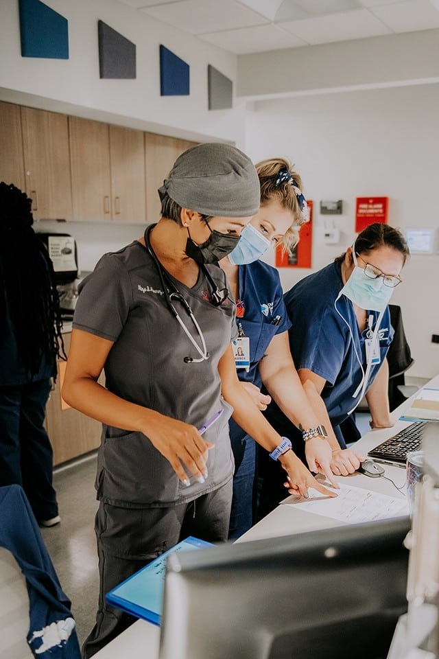
Cardiac catheterization can help your provider diagnose blockages in the coronary arteries that may cause heart attacks. Cardiac catheterization is also used to measure certain pressures and flows in the heart which are helpful to diagnose other conditions. Cardiac catheterization studies are invasive procedures used to examine structures in the heart, typically coronary arteries, the aorta, pulmonary vessels, and cardiac chambers. Images of these structures are obtained by inserting a catheter (similar to a very small tube) into vessels found in the arm or leg, and tracing the pathway with a contrast dye. Most of these procedures can be competed in our outpatient surgical center, but sometimes they are performed in a hospital’s cardiac catheterization lab. You will be asked to fast for 4 to 6 hours prior to the diagnostic exam. You will be given intravenous conscious sedation that allows you to remain awake yet relaxed during the procedure. Afterwards, you will be taken to a recovery area to be monitored until your provider determines it is safe to discharge you. If this procedure is necessary for you, your provider will discuss the risks and benefits prior to the procedure.

Left Heart Catheterization

This procedure is used to determine if an individual has significant coronary artery disease. The test provides advanced imaging of the arteries that feed blood to your heart to determine if there are blockages that restrict blood flow.

This test is used to measure pressures in the left side of the heart to determine left ventricular performance. This test can also diagnose conditions such as congenital coronary artery abnormalities and myocardial bridging.

Right Heart Catheterization

A right heart catheterization is a diagnostic procedure used to measure pressures in the pulmonary artery and right side of your heart.

This type of catheterization is not routinely performed during the cardiac catheterization procedure. However, when symptoms or other conditions call for further investigation (for example: unexplained shortness of breath, valve disease, pulmonary hypertension), your provider may order a right heart catheterization.

The renal arteries are your body’s main blood supply to your kidneys. Abnormalities in the renal artery can cause other problems such as high blood pressure and renal insufficiency. Renal artery imaging provides detailed images of the renal artery and blood supply and assesses the vessels blockages, narrowing, or bulging. If your doctor suspects there may be disruption in the function of the renal arteries, renal artery imaging may be indicated. Images of the renal artery can be captured via ultrasound, CT scan or renal angiogram, much like the other contrast dye imaging for other vessels.

Renal Artery Imaging

Implantable Cardiac Monitor

Loop Recorder
A loop recorder is an implantable cardiac device used to monitor your heart’s rate and rhythm and is useful if your doctor suspects a disruption in your heart’s electrical system. Your provider may order this monitor for you if you infrequently have symptoms such as palpitations (feeling like your heart is racing or skipping a beat), or signs and symptoms of a stroke with no known cause. The loop recorder insertion is a simple procedure performed in our outpatient surgical center. A local anesthetic is used to numb the left chest area where the paper clip-sized monitor is inserted so that you do not feel any pain. The monitor is then inserted, and surgical glue is applied. With a battery life of up to three years, the monitor is designed to provide data over an extended period for the most accurate information. Your provider will routinely monitor your device for arrhythmias as they see necessary.
Sometimes a heart may not keep up the pace with the needs of the person, and their heart isn’t beating quickly enough. A pacemaker is a small device inserted into the chest to treat certain cardiac arrythmias caused by a disruption in the heart’s electrical current. The pacemaker automatically signals your heart to beat properly if your heart is beating too slowly. Whether in our outpatient surgical center or in the hospital, before the procedure you are given medication to help you relax. A small incision is made in your left chest, and the pacemaker lead is inserted into the chamber in your heart needing stimulation. The lead is connected to a generator that is then inserted in your chest wall, and your incision is closed. Your vital signs are monitored during the procedure. After the procedure, you will be taken to a recovery area where your vitals will continue to be monitored, and the new device is verified to be working properly. Once discharged, you will need to have someone drive you home, and you will also need to adhere to postoperative instructions given to you by your doctor. If this procedure is necessary, your provider will discuss the risks and benefits prior to the procedure.

Pacemaker Implants

Additional Services Provided

Coronary Artery Imaging

Heart catheterization for congenital anomalies

Scroll to Top

How can we help?